Even better operations with LED photoacoustic imaging
Item 1
of
1
Technology is helping the medical world progress and it is an important motivating factor for Saskia van Heumen, who graduated cum laude at the end of 2021 from the Faculty of ME. Her research into using LED photoacoustics in medical imaging brought the medical world a step further. It earned her the title Best Graduate of her Faculty.
With a mother who was a doctor and a father who was a software engineer, Saskia van Heumen’s choice of studying Clinical Technology at TU Delft meant that the apple did not fall far from the tree. “I always loved hearing my mother’s stories about her fellowships,” she says. “I was fascinated by the human body, but a profession as a doctor did not appeal to me. And as I also love technology, I went looking for applied technology in the medical field. I found out about Clinical Technology’s open day and this turned out to be the perfect study for me.”
Medical imaging
Saskia was in the first cohort of this programme that started in 2014. After her bachelor, she went to England for a year to do a post graduate at University College London on medical physics and imaging. “I liked it so much that once I was back in the Netherlands, I started the Technical Medicine - Imaging & Intervention master’s. It’s an alliance between TU Delft, Erasmus MC and LUMC. You do four internships of 10 weeks in the hospital in the second year of the master’s. I already had quite a lot of theoretical knowledge about medical imaging and was looking forward to putting it into practice. I was able to do various bits of research into the use of images in medical interventions in several hospitals. One of these was during my internship in dental, oral and maxillofacial surgery. I helped make surgical 3D moulds from CT scans, of the mouth for example. Surgeons use these moulds so they know exactly how to operate. I found this incredible as you immediately see how your research helps the quality of healthcare.”
From sound to light
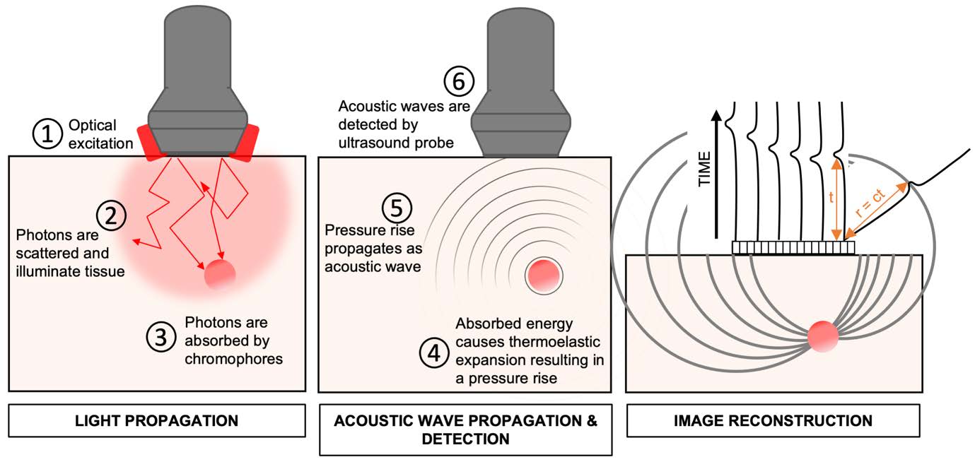
During her graduation internship, Saskia learned about photoacoustic imaging. Make images of the body was a relatively new and very promising technology. “We already know about ultrasound machines which use sound waves to create images,” Saskia explains, “but in photoacoustic you send light pulses into the body instead of sound waves. The LED light is absorbed and this ensures that the tissue warms up locally and expands. This then creates sound waves. As different types of tissue and injected contrast medium absorb light differently, you can use different wave lengths to create images and distinguish them from each other. Together, the sound waves that result then form an image.”
Lymphedema
One of the potential applications for photoacoustics with LED is imaging damaged lymph vessels. Some breast cancer patients develop serious lymphedema (fluid accumulation) in their arms from the treatment. It is very unpleasant as it limits the use of the arm and affects the quality of life. Surgeons can help lymphedema by carrying out a complicated operation in which they make a sort of bypass in the finely meshed vascular system. “This is now done using another proven technique called near-infrared fluorescence (NIRF) imaging. But this technique does not show exactly how deep the lymph vessels are and exactly where the blood vessels are. I examined if LED photoacoustic imaging would reflect these structures more precisely in my graduation project, the Photo-acoustic Imaging of the Lymphatic Vessels project.
I examined if LED photoacoustic imaging would reflect the structures of lymph and blood vessels more precisely in my graduation project.
Saskia van Heumen
Best Graduate 2022 of the faculteit of ME
I examined if LED photoacoustic imaging would reflect the structures of lymph and blood vessels more precisely in my graduation project.
Saskia van Heumen
Best Graduate 2022 of the faculteit of ME
Medical ethical check
Saskia did her research in practice. “I wanted to do a clinical study as part of my graduation project. I could then find out if photoacoustic imaging really would prove useful in these complicated bypass operations.” To do medical scientific research with people, you need the agreement of the Medisch-Ethische Toetsingscommissie (medical ethical committee, METC). Saskia wrote a research proposal for this purpose. “It was hard as a new regulation had just been passed governing the use of medical equipment in clinical trials and nobody had any experience with it yet. So I had to do a lot of work like phoning the manufacturer of the equipment and so on. It was nerve wracking getting my research proposal through this process.”
Patients
“So it was really amazing after all the, sometimes frustrating, preparations to finally start doing the real work with patients. The patients that took part in the research project first went to the plastic surgeon for the regular examination and then came to me for the photoacoustics. This was done on the condition that the images would not affect the decisions of the surgeon.” Saskia is proud of the outcome. “My research was the first step in showing that LED-based photoacoustics could map the lymph vessels and blood vessels in greater detail than the technique used now.”
Organised go-getter
The Board of Examiners praises Saskia for her versatility, determination and her organised way of working. She herself remains modest and stresses that she received a lot of support from her thesis supervisor Professor Gijs van Soest. “There was already a lot of knowledge about photoacoustics in the research group, especially about technical medical care and Jonas Riksen, a doctoral candidate, and I could brainstorm well. Dalibor Vasilic, the plastic surgeon who does these lymph vessel operations, helped me a lot too. He allowed me to shadow him so that I would know exactly what kind of information he needed for his operation plan and how the medical decision-making process went.”
Future
Since then, Saskia has had her systematic review published in Annals of Surgical Oncology, and her clinical study is under review for publication in Photoacoustics. She will start her doctoral research at the Amsterdam UMC in January 2023. Will it be on photoacoustics? “No,” she laughs. “My interest in imaging technology is wider than that. I will look at the diagnostics of lung cancer using bronchoscopy, a tube with a camera attached to the end which helps you see the lungs from the inside. I will help assess the use of new imaging methods to help doctors make better diagnoses.”
I will help assess the use of new imaging methods to help doctors make better diagnoses of lung cancer.