
The formation of kidney stones on a microscale
Some factors that influence the formation of kidney stones are known, but little is known about the exact physio-mechanical processes that take place within the kidney. Researchers from TU Delft developed a method to watch the formation of kidney stones on a microscale, in a so called microfluidic platform. By slightly adjusting the pH and the concentration of specific minerals, the formation could be slowed down or inhibited completely. The research is now published in Biomicrofluidics.
Kidney on a chip
About 12% of the world population suffers from kidney stones and the incidence of the condition is increasing worldwide. In many cases, patients suffer from re-emerging stones. Therefore, it is important to precisely understand the underlying processes of the formation of kidney stones. “The kidney still is a sort of black box”, says researcher Burak Eral, “we can measure what goes in and what goes out, but it is hard to really look inside.” The researchers developed a droplet-based microfluidic platform, a so called ‘kidney on a chip’, in order to see kidney stone formation on a microscale.
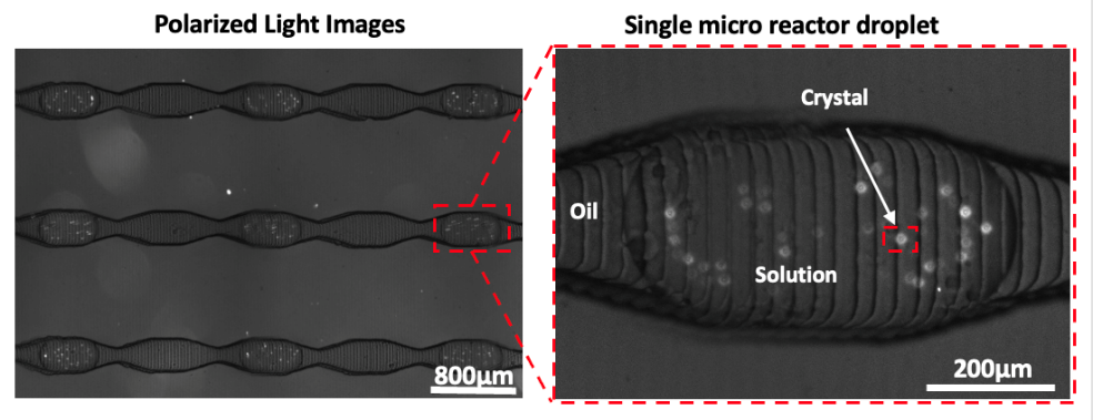
The kidney on a chip basically consists of a small tube and two inputs: fluid that resembles kidney fluid, and oil. After a drop of kidney fluid is pumped into the channel, a drop of oil is added. In this way, the droplets with kidney fluid are separated from each other, forming microscopic kidney environments. The tube and the droplets are so small, that even the tiniest formation of a kidney stone can be observed with a microscope.
Artificial urine
By adjusting the composition of the kidney fluid, the researchers were able to observe exactly which factors influence the formation of kidney stones. Changes in the pH-level and in the concentration of Magnesium caused the formation to be slowed down, or even inhibited. Researcher Fatma Ibis: “It’s the first time that this kind of experiment is done at such a small scale and also allows to observe emerging of crystals simultaneously. In larger scales, it’s only possible to watch the growth of already formed kidney stones. Now, we can really see direct effects of changes in the kidney fluid.”

The researchers only used essential ingredients to resemble the kidney fluid, in reality kidney fluid is much more complex. Eral: “It is the beginning of a long journey. A next step would be to use artificial urine in our kidney on chip and make the system more complex.” Finally, the researchers think it will be possible to use the mechanism with the urine of patients, to determine a specific treatment for their condition. Ibis: “Many factors play a role in the formation of kidney stones, and the factors differ for each patient. Personalised treatment could be a very efficient way to help those patients.”
More information
Ibis, F., Yu, T. W., Penha, F. M., Ganguly, D., Nuhu, M. A., van der Heijden, A. E. D. M., Kramer, H. J. M., & Eral, H. B. (2021). Nucleation kinetics of calcium oxalate monohydrate as a function of pH, magnesium, and osteopontin concentration quantified with droplet microfluidics. In Biomicrofluidics (Vol. 15, Issue 6, p. 064103). AIP Publishing. https://doi.org/10.1063/5.0063714
Contact
Burak Eral, H.B.Eral@tudelft.nl, +31 15 27 86715
Fatma Ibis, F.Ibis-1@tudelft.nl, +31 15 27 86658
Dimmy van Ruiten, press officer, D.M.vanRuiten@tudelft.nl, +31 15 27 81588

