Cytoskeletal Crosstalk
Quantitative imaging of cytoskeletal crosstalk at the nanoscale
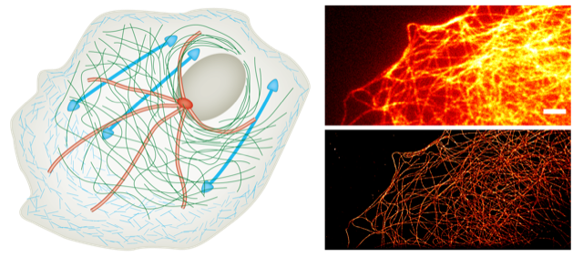
What? Master project to establish new multicolor super-resolution imaging to unveil the nanoscopic cytoskeletal structures (actin, microtubules and intermediate filaments) of cells as well as their interaction (crosstalk) and relate it to their active mechanical behaviour. You will optimize multicolor imaging and focus on quantitative network analysis of multicolor super-resolved networks at the nanoscale or on extending imaging from 2D to 3D. At a later stage, we will investigate how different surface micropatterning influences cytoskeletal crosstalk. This is a joint Kavli Institute for Nanotechnology Delft funded project between the Koenderink Lab and the Grussmayer Lab @BN/AS.
Starting date & contact: available immediately; Kristin Grußmayer, Email: k.s.grussmayer@tudelft.nl , Gijsje Koenderink, Email: G.H.Koenderink@tudelft.nl
For you? You learn: Cell culture and labeling, state-of-the-art super-resolution microscopy methods and advanced image analysis as well as visualization and presentation of experimental data. Depending on your background, the project may include micropatterning of surfaces for controlled cell adhesion. Your background: nanobiology, (bio-) physics/engineering/chemistry or related.