Marlies Goorden
Nuclear medical imaging techniques, such as PET and SPECT, utilise radionuclide tracers (containing unstable atoms that emit radiation when decaying) to provide unique information on tissue function and biologic processes. Rising star Marlies Goorden develops algorithms, models, and novel geometries to continually enhance their imaging properties.
The medical imaging techniques we develop oftentimes make it into (pre-)clinical practice
For the first few years after graduating from TU Delft as a physicist, Marlies Goorden explored theoretical solid-state physics. ‘I found it highly interesting,’ she says. ‘But in the end, it often wasn’t even possible to really test a theory I developed.’ Now, as an assistant professor in the Biomedical Imaging group of Professor Beekman, she very much enjoys the medical aspect of her work. ‘I still do a lot of theoretical work but the techniques I and my colleagues develop oftentimes make it into (pre-)clinical practice, thanks to a close collaboration with MILabs BV. The ongoing convergence between TU Delft, Erasmus MC, and Erasmus University narrows the gap between engineers and medical specialists even further.’
Breast imaging
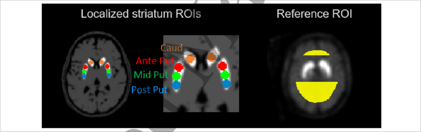
A few years ago, as part of her Vidi grant from the Dutch Research Council (NWO), she built a prototype breast SPECT scanner (Single Photon Emission Computed Tomography). The national breast cancer screening program uses two-dimensional X-ray images. Follow-up imaging is indicated if potential abnormalities are detected. ‘Using radiotracers, and a SPECT-camera consisting of many pinholes used over multiple imaging angles, we aimed for this follow-up examination to yield high-resolution 3D images,’ Marlies says. ‘The goal was to improve the distinction between tumour and healthy tissue. Thanks to the wide variety of available radionuclide tracers, such a scan could also reveal certain tumour characteristics. Ultimately, perhaps, this could make a biopsy unnecessary for determining the best course of treatment or for monitoring therapy.’
Using a SPECT-camera consisting of many pinholes, we can obtain resolutions close to the thickness of a human hair.
A nuclear microscope
Although the prototype breast scanner didn’t make it into routine clinical use, the idea behind it is still very much alive. This summer, Marlies was co-awarded a grant through the Convergence Health & Technology Open Mind Call in which she proposed to realise a high-resolution nuclear microscope based on the breast scanner technology. Marlies: ‘We collaborate with Julie Nonnekens of the Radionuclide Therapy group of Erasmus MC. They develop novel radiopharmaceuticals and want to quantitatively assess their diffusion in tumour tissue. Our simulations have shown that we may be able to obtain a resolution of less than one-twentieth of a millimetre – the width of a human hair.’ She has various other grant proposals pending, both national (NWA, Dutch Research Agenda) and within Convergence.
Advanced reconstruction algorithms
Speeding and improving image reconstruction are an integral part of all of Marlies’ projects. ‘In clinical practice, users often do not apply optimal image acquisition parameters as it may lead to prolonged image reconstruction times,’ she says. It is another area of research in which she likes to operate at the cutting edge of technology. Together with a PhD student, she also applied neural networks to improve the so-called attenuation correction for SPECT imaging – a method to correct for the loss of radiation due to scattering and absorption within the person being imaged. ‘When imaging small animals or slices of tissue, you don’t have to apply such a correction,’ she explains. ‘But in patients it can lead to artifacts and, therefore, uncertainty in the location and shape of a lesion.’ Last but not least, she is collaborating with Dennis Schaart of the TU Delft Medical Physics & Technology group. Their combined aim is to improve the quality of the existing cone-beam CT at the brand new HollandPTC proton therapy centre at TU Delft campus.
In clinical practice, optimal image reconstruction may lead to prolonged reconstruction times
Keep up with Marlies
So, pretty clear who you should contact if you are in need of a state-of-the-art (high-resolution) radiation-based medical imaging solution. Simply give Marlies a call or join her during one of her many runs.