Sample preparation
Remember- you are imaging a physical interaction between light and the chosen label for your target. Even the most sophisticated microscope will only give you good/reliable enough data as your sample quality!
After phrasing your question, and before diving into tedious imaging sessions and analysis optimizations, take the time to think about your sample preparation:
Choice of labelling:
- Affinity, Size – Consider the effect on the interaction with your target.
- Fluorescent Proteins/Fluorescent Dyes
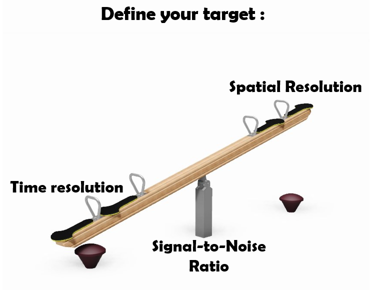
- Spectral properties: aim for good signal to noise ratio, avoid crosstalk for co-localization.
- For long-term imaging: consider phototoxicity and photobleaching
- Are you interested in Correlative light-electron microscopy? Make sure your sample label fits both.
Boosting sample resolution:
- Clearing
- Expansion Microscopy- a bench strategy for Super-resolution
Including controls for quantitative measurements
- Find the center using fluorescent beads for co-localization and deconvolution
- Always use a single label for multi-label imaging
- Try to also include a biological control to verify your observation
- Check antibody specifity with no primary antibody control!
- Statistics Statistics Statistics!!