Liquid-phase Electron Microscopy for microtubule dynamics
Nemo Andrea
Collaborators: Project shared between Dogterom lab and Schneider lab (Leiden University, LIC, SBC)
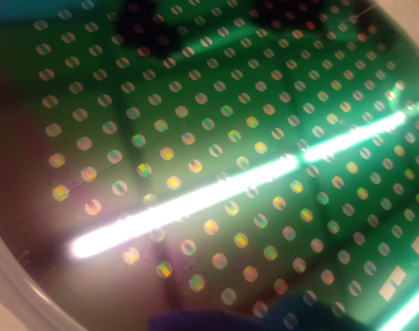
Cryo-EM studies have given us ever better glimpses of the microtubule end during growth, but the fundamentally static nature of vitrified systems combined with the heterogeneous nature of the microtubule tip prevent a complete understanding of this dynamic area. To address this issue, we are investigating the system with liquid-phase EM, where the system remains in the liquid state, trapped between thin electron-transparent windows. We use both conventional ‘veil-type’ graphene liquid cells and specially developed microfabricated flowcells with the aim to observe dynamics at the nanoscale in TEM.
