
Over 250 million people have been infected with schistosomiasis a waterborne parasitic infection. Early treatment is key in preventing deaths and reducing long term morbidity. To ensure this, access to accurate diagnosis, which is frequently lacking in rural Africa, is crucial. We aim to improve the quality of and access to diagnosis of Schistosomiasis in rural Africa with the Schistoscope. The device will be based on smart algorithms used with an integrated Android mobile and feature locally manufacturable embodiment. The aim of this project is to develop a low cost, a smart diagnostic tool that can be locally manufactured and repaired to be accessible to the last mile health care providers. The Schistoscope will be designed to not require high expertise, thereby increasing the accessibility of the device.
Open Mind Schistoscope
The Schistoscope
Challenges
Early treatment of Schitosomiasis critically reduces the risks, for which access to accurate diagnosis is crucial. The diagnostic tools recommended by WHO have shortfalls. Either they are expensive or availability is subject to supply and some are also not deployable in a field setting. Diagnosis is usually performed by light microscopy (expensive and maintenance challenges) which involves trained medical staff who are scarce as well as can cause human-error.
Schistoscope
We propose a Schistoscope - which offers an integrated diagnostics solution (sample preparation and diagnosis) with the support of a smart algorithm (for detection and quantification of the Schistosoma eggs) which can be produced and maintained in Africa (use of locally available materials and manufacturing process). We will explore open source possibilities to improve accessibility and local manufacturability such as integrating 3D printing possibilities in the universities and maker labs. The core of the Schistoscope is a smart imaging platform composed of a locally available low-cost Android device, a low-cost imaging lens, and image recognition and classification algorithm. For sample preparation, 10 ml of urine will be dropped using a syringe through locally available filter material which is positioned on a plastic sample holder. The sample holder and the smartphone will be inserted in an ‘integration box’ to position the camera lens exactly above the sample. By capturing one image (one field of view) the algorithm will be able to diagnose the sample and share the results directly for treatment of the patient.
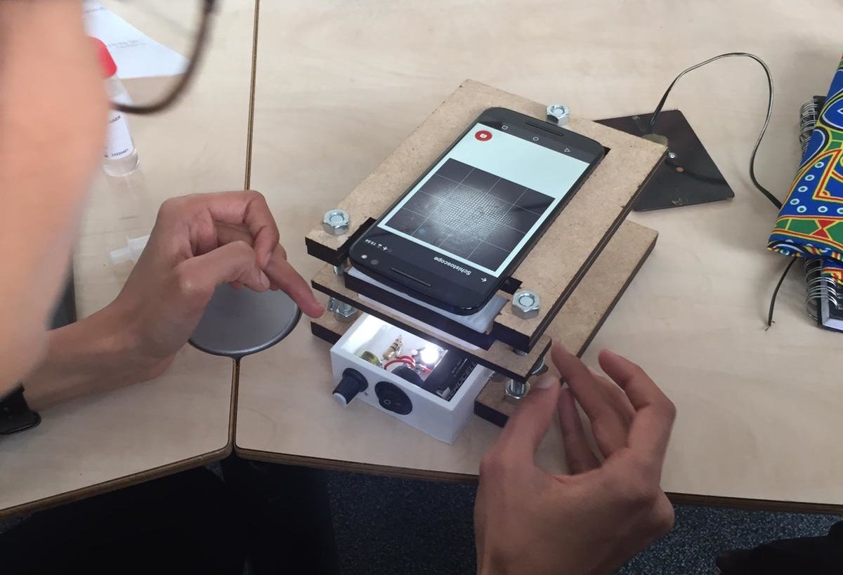
Current stage of the project
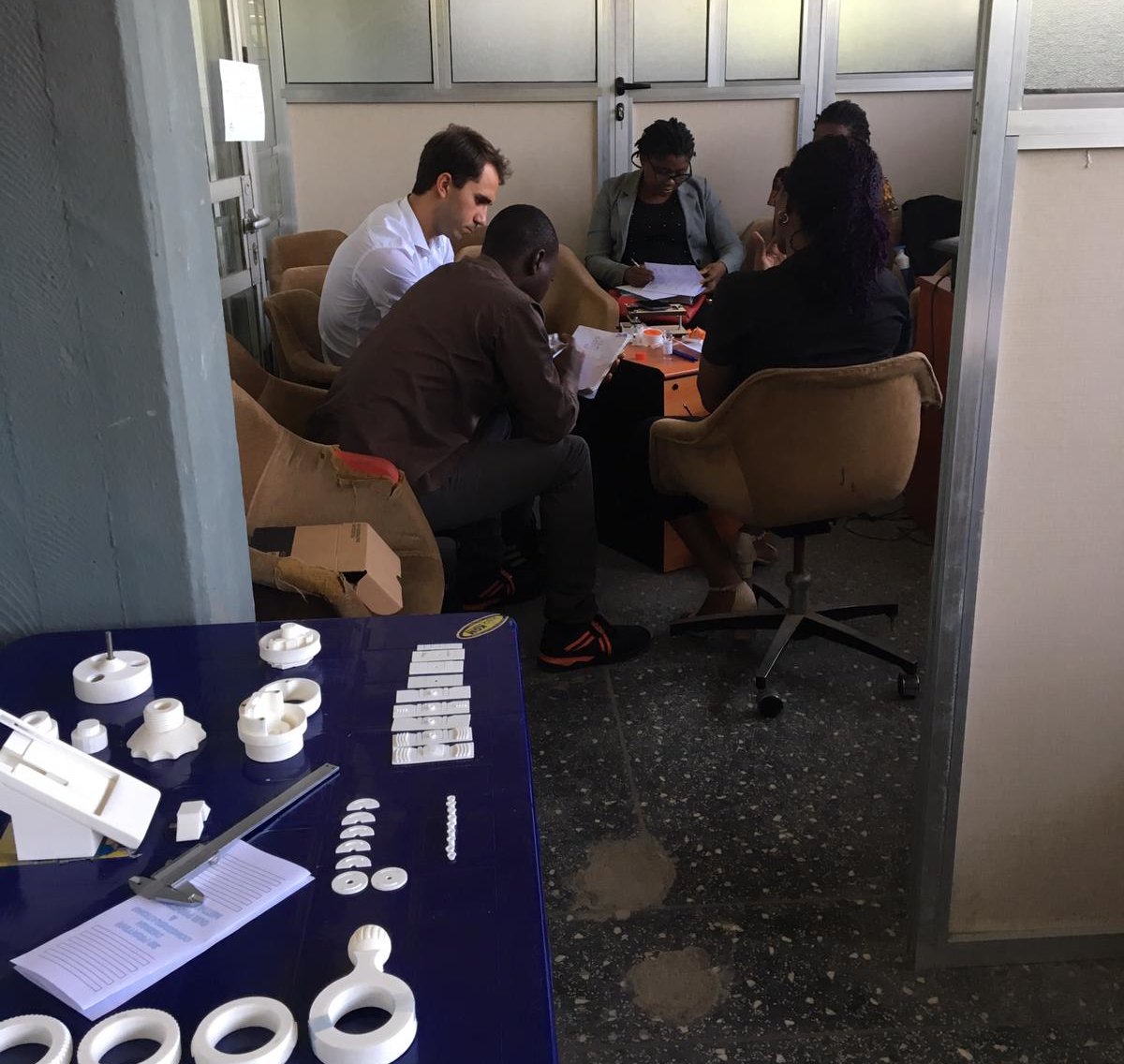
The student team (MSc Industrial Design Engineering and System and Control Engineering, TU Delft) is dedicated to developing the technically feasible prototype of the concept. A simple mock-up was made based on the initial proof of concept. During the first visit to Nigeria, it has stimulated the discussion among the interdisciplinary partners and collaboration for an optimal solution.
Contributions
Delft Centre for Systems and Control l Delft University of Technology
Department of Parasitology l Leiden University Medical Centre
Industrial Design Engineering l Delft University of Technology
Delft Global Initiative l Delft University of Technology
Public Health l University of Ibadan, Nigeria
College of Medicine l University of Lagos, Nigeria

Dr. Ir. Jan-Carel Diehl
- +31 (0)15 27 89729
- J.C.Diehl@tudelft.nl
-
Room B-3-350
Landbergstraat 15,
Delft University of Technology
