Bringing the medical world into science non-fiction
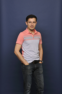
Zaid Al-Ars, Associate Professor in the Computer Engineering Lab at TU Delft, is a computer engineer who possesses a pretty decent internal hard drive himself. He studied electrical engineering and obtained a doctorate in computer technology before joining first-year students again to study medicine. He is one of the first engineers who are sincerely fascinated by human beings. And he has the drive to introduce ‘Star Trek’ technology into a very conservative world.
Cutting people open is outdated
Zaid Al-Ars

You say that the medical world is still stuck in the nineties. Why is it that the medical world remains so far behind technology?
“The medical world is very conservative. That is not so surprising: it deals with living things, so you cannot be too cautious. Medical specialists want to understand what they are doing as well as possible. New technology can be baffling and specialists feel it is safer to use their trusted instruments. Fortunately, this same world is gradually realising that new technology can help doctors enormously in making diagnoses, providing support for interventions and improving the results of such interventions. I can see technology departments appearing in hospitals in which engineers, ICT specialists and other non-medical specialists are increasingly working in collaboration with doctors. That is something that we can be proud of having brought about in the medical world.”
That will not have been easy?
“No. Years ago, when I decided I wanted to work in this world, I attended lectures in Leiden. I didn’t just read a bunch of 1000-page biology books, I really wanted to understand the material. I also talked to researchers, biologists, doctors, and so on. I had already devised a new implementation of an algorithm that is widely used in the medical world, which yielded a much better result. At least in theory it did… I told my professor in Leiden about it enthusiastically. However, he then asked me: ‘What is the biological significance of your results?’ And of course that was a question I couldn’t answer. My professor then continued: ‘My algorithm may be less accurate, but I understand the result. I can actually use it. Every body is different, which makes accuracy less important.’ Then I realised that, if you want to connect two worlds, you need to understand each other’s way of thinking. My biology professor in Leiden ultimately became one of my first research partners. ‘No other researcher tried so hard to follow my lectures’, he told me later. I worked with professor Herman Spaink in Leiden for many years and developed one of my first implementations together with him.”
How did you come to decide that you wanted to go in that direction?
“That was after getting my PhD. I had a post-doctoral position in Delft and wanted to broaden my horizons. At the time there was revolution going on in biology, related to the rapid developments in genetics. I found it extremely interesting to learn about the basic building blocks of life. And it also enabled me to help people directly, which was something I missed in technology. If you can apply technology in the medical world, you will have a direct impact on society. In retrospect, I made a very good choice.”
Then I realised that, if you want to connect two worlds, you need to understand each other’s way of thinking
Fast forward to now. One of your projects focuses on non-invasive surgeries. Is that the future?
“Actually, cutting people open is outdated. There is a movement in the medical world that strives to be as non-invasive as possible, while still enabling you to make a diagnosis and carry out interventions. We are making significant progress. And we are working on this with Philips, who develops medical imaging equipment.
“Together, we want to turn a science fiction-like scenario into reality, where a patient lies in a type of diagnostic imaging machine. A specialist can then view the patient’s hard tissue as well as his soft tissue. For this, you need a variety of devices and different types of radiation to identify and image it. Once you have all the body images, you can merge them in real time. This is called image fusion. And all this is done in 3D. If doctors then want to carry out an intervention – an operation – they will also want to know precisely where in the patient lying in their operating theatre this must be done. So you add an image of the ‘exterior’ of the patient and all of a sudden you have a complete inside-out image of a person on the screen that you can rotate, zoom in on and manipulate. The organs also move on the screen in real time. A doctor can then enter the body at exactly the right spot with an instrument to begin treatment. Incidentally, those instruments are also developed here in Delft. They are designed so that you no longer have to enter the body in a straight line. You can manoeuvre around an organ to reach a certain point, without touching anything you want to avoid.”
All of a sudden you have a complete inside-out image of a person on the screen that you can rotate, zoom in on and manipulate
So it’s like a game of Operation, only the Star Trek edition?
<Laughs > “Yes, it is all very futuristic. Unfortunately, it is not as easy as an old-fashioned game of Operation. It is very difficult to merge images from different sources as well as incorporating all those real-time dynamic images of the organs. We have to render a huge amount of information very quickly on the screen, which is also of very good quality. What you see is what happens.”
The DNA of Zaid Al-Ars
Zaid Al-Ars is an Associate Professor in the Computer Engineering Lab at TU Delft and heads the research into big-data systems. He is working on bottlenecks in the scalability of big-data applications in multicore architectures. He is best known to the general public for his work on DNA sequencing, which resulted in the start-up Bluebee, where cloud computing, high-performance computing and genomics applications come together. Two months ago, he and his research group applied for another patent on DNA analysis. This new technology shortens the process of DNA sequencing for clinical diagnosis from a few weeks to just a couple of hours.
Text & Photo: Marieke Roggeveen | April 2017