Major Areas of Research
The section Biomedical Imaging has a focus on nuclear imaging techniques that utilize radionuclides (i.e. PET and SPECT) to provide unique information on tissue function and biologic processes. We actively work on various aspects of scanner development; we develop analytical scanner models and perform computer simulations to optimize scanners, we construct novel collimators for nuclear tracer imaging, we develop gamma detectors for PET and SPECT and we work on highly accurate and fast iterative tomographic image reconstruction. We also work on unique hybrid nuclear imaging (simultaneous PET-SPECT combined with e.g. high performance X-ray CT and optical tomography). Research is carried out in close collaboration with academic medical centers and industry.
Preclinical imaging systems
In the past 15 years, our group has developed a complete new line of ultra-high resolution SPECT-PET systems for small laboratory animals (U-SPECT with 0.25 mm resolution, VECTor PET-SPECT with 0.4 mm resolution SPECT simultaneously with 0.6 mm resolution PET and 0.14 mm resolution stand-alone 3D autoradiography). These high resolutions are attained by unique physical collimation methods, algorithms and software such as advanced image reconstruction, smart robotics and new scanner system geometries. Many of these technologies are now in use in biomedical research labs world-wide often incorporated in hybrid systems that allow to combine e.g. PET, SPECT, CT and Optical Tomography. Current research not only focuses on improving spatial and temporal resolution for current scanners but also on expanding the range and combinations of isotopes that we can image. Generally we are aiming to maximize quantitative accuracy of 3D and 4D images for virtually any available optical, gamma, positron or co-emitting tracer molecule or tracer combination in the 30 keV-1 MeV energy range.
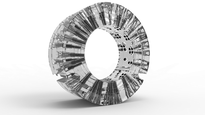
Dedicated collimator to image SPECT and PET tracers simultaneously (Goorden et al, VECTor: A Preclinical Imaging System for Simultaneous Submillimeter SPECT and PET, JNM 2013).
Clinical imaging systems
Advances in preclinical SPECT instrumentation such as the application of novel pinhole collimation, image reconstruction and new detector technologies, are beginning to carry over into clinical imaging. Our group is highly involved in improving clinical SPECT with support of e.g. a joint project with NWO-FOM and industry. We are actively involved in the G-SPECT project (Innovation of the Year, 2015 World Molecular Imaging Society) to achieve <3 mm resolution in patients. Additionally, we are constructing a new prototype Molecular Breast Tomosynthesis system for imaging SPECT tracers to e.g. detect and characterize tumors.
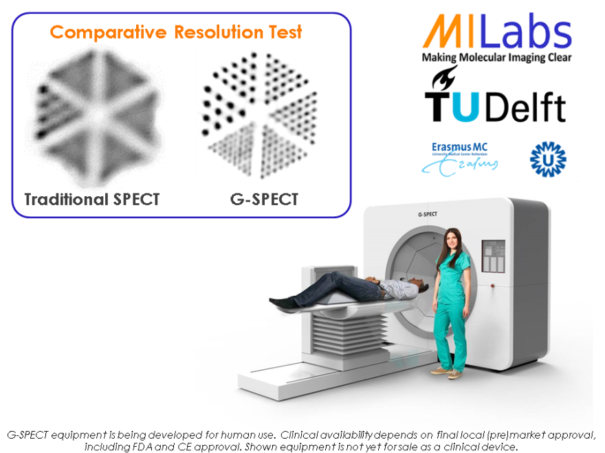
G-SPECT system for high-resolution multi-pinhole clinical SPECT.
Gamma ray detectors
A key part of any nuclear imaging technology is the gamma ray detector, which should provide the position and energy of an incoming gamma photon. We have worked on the optimization of gamma ray detectors by using various scintillating materials, new combinations of sensors and unusual light guide geometries as well as solid state gamma detectors. As we develop our own high-speed electronics for read-out, we have the flexibility to incorporate our own detection algorithms to better estimate the gamma photon’s entry point.
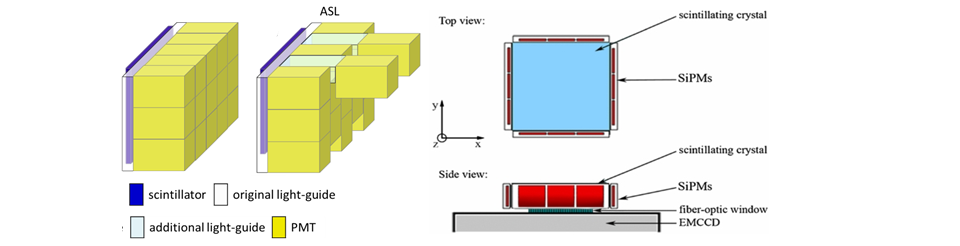
Different types of novel gamma ray detectors developed in our group. Left: dedicated PMT-lightguide geometry for breast scanner. Right: EMCCD gamma camera with side detection (Salvador et al, PMB 2012).
Image reconstruction
Ideally, image reconstruction is based on accurately modelling the gamma photon transport through the whole system, including the animal/ patient that is imaged, the collimator and the gamma detector. We develop methods for fast and accurate photon transport modelling. These can be based on measured point source responses combined with smart interpolation or full scanner simulations, e.g. based on ray tracing or Monte Carlo methods. These photon transport models are incorporated in statistical iterative image reconstruction. As these iterative methods are notoriously slow, we have also introduced several algorithms that allow to accelerate reconstruction by orders of magnitude. Especially for the new clinical systems that we are developing, attenuation and scatter in the patients play a large role and we are therefore working on new methods to accurately correct for these effects in order to arrive at quantitative images that accurately reflect the radioactivity concentration.