Shining light on densely interwoven nerve fibres inside the brain
Disentangling the complex nerve fibre network of the brain is becoming easily accessible with scattered light imaging (SLI): researchers in Delft, Jülich (Germany) and Stanford (USA) successfully combined light and X-ray scattering with MRI to discern nerve fibre trajectories, also in regions with highly entangled fibres. SLI revealed the trajectories at highest detail, while being significantly faster and cheaper than X-ray and MRI techniques. Such detailed mapping is essential for a better understanding of how nerve fibres are wired inside the brain. The study was published in the journal eLife.
Pathways in the brain
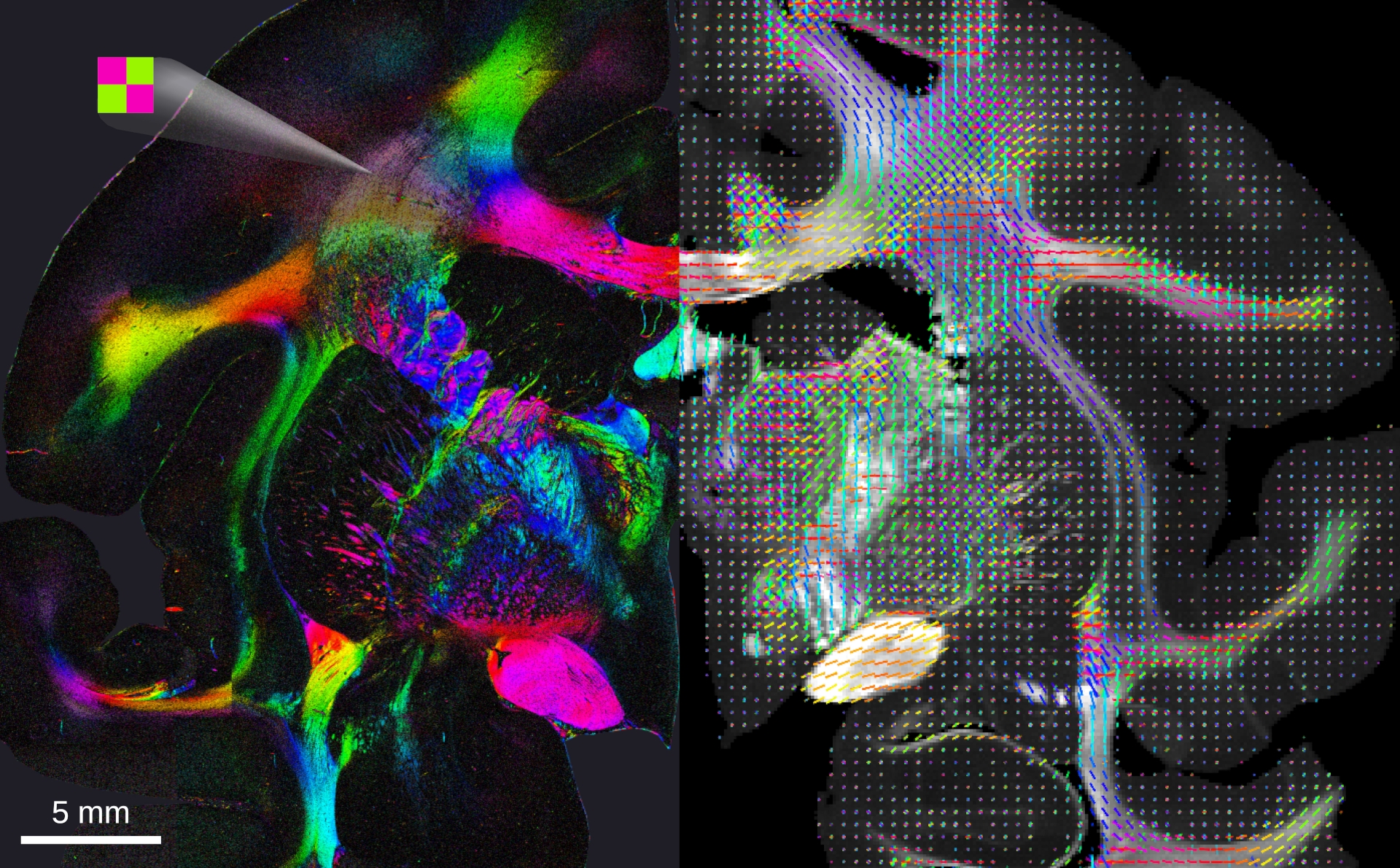
The different areas in the brain are connected to each other via billions of nerve fibres. These connections are vital for proper brain function. The quest for a comprehensive map of all neural connections critically depends on imaging techniques that can disentangle these fibres, most of them only about a micrometre thin. Particularly challenging are regions with densely packed and highly interwoven nerve fibres. Miriam Menzel, Assistant Professor at the Department of Imaging Physics of TU Delft, developed the SLI technique to study such fibre constellations: “We shine light under different angles through hair-thin brain slices and analyse the resulting scattering patterns. We’re not taking a picture of neurons or synapses; we want to know how they are wired. This is important for understanding brain function and dysfunction.”
More accessible, cheaper and faster
Small-angle X-ray scattering (SAXS) is an established method in material science to look into how different structures are organised with a synchrotron, while diffusion magnetic resonance imaging (dMRI) is an important technique in clinics for visualising the three-dimensional nerve fibre network of the brain. “We have now shown that SLI data are consistent with those from SAXS and dMRI in the examined brain slices, but SLI provides higher resolution than dMRI and is more accessible, cheaper and faster than the other techniques. This is an important milestone,” Menzel says. “We can perform SLI measurements with a simple LED light source and camera in just a few seconds, requiring neither a multi-million synchrotron nor an MRI scanner. As portable system, it could easily be set up in pathology laboratories to assist clinical research.”
Microscopic resolution
Menzel has spent the past few years working on the SLI technique, first in Jülich and now in Delft. She also implemented it in Stanford, where her fellow researchers performed SAXS and dMRI measurements on brain samples also imaged with SLI. “Most imaging techniques struggle to discern individual pathways in dense brain structures containing many entangled or interwoven nerve fibres,” Menzel explains. “SLI provided fibre orientation maps with microscopic resolution in these dense regions.” Especially the two-dimensional (“in-plane”) fibre orientations were discerned with high precision.
Next steps
“Being in Delft brings exciting opportunities to develop the technique further and work on new applications,” says Menzel. The team plans to also apply SLI to other types of fibres, such as muscle and collagen fibres, and to enlarge the tissue area that can be studied. The aim is to develop a small and portable system that can easily be deployed in other labs. “In the long term, we hope to apply the technique in clinics as well.”
More information
Using light and X-ray scattering to untangle complex neuronal orientations and validate diffusion MRI. Miriam Menzel, David Gräßel, Ivan Rajkovic, Michael M Zeineh, Marios Georgiadis. eLife 12:84024 (2023) DOI: https://doi.org/10.7554/eLife.84024
