Towards single-cell biopsy with 3D printing
Murali Ghatkesar, assistant professor in the Department of Precision and Microsystems Engineering, has developed a new method through 3D printing that makes it easier, quicker and more efficient to perform single-cell biopsies. It is the first time that 3D printing is being used for the production of micro- and nanofluidic equipment. The results of his research were published in the scientific journal Lab on a Chip this month.

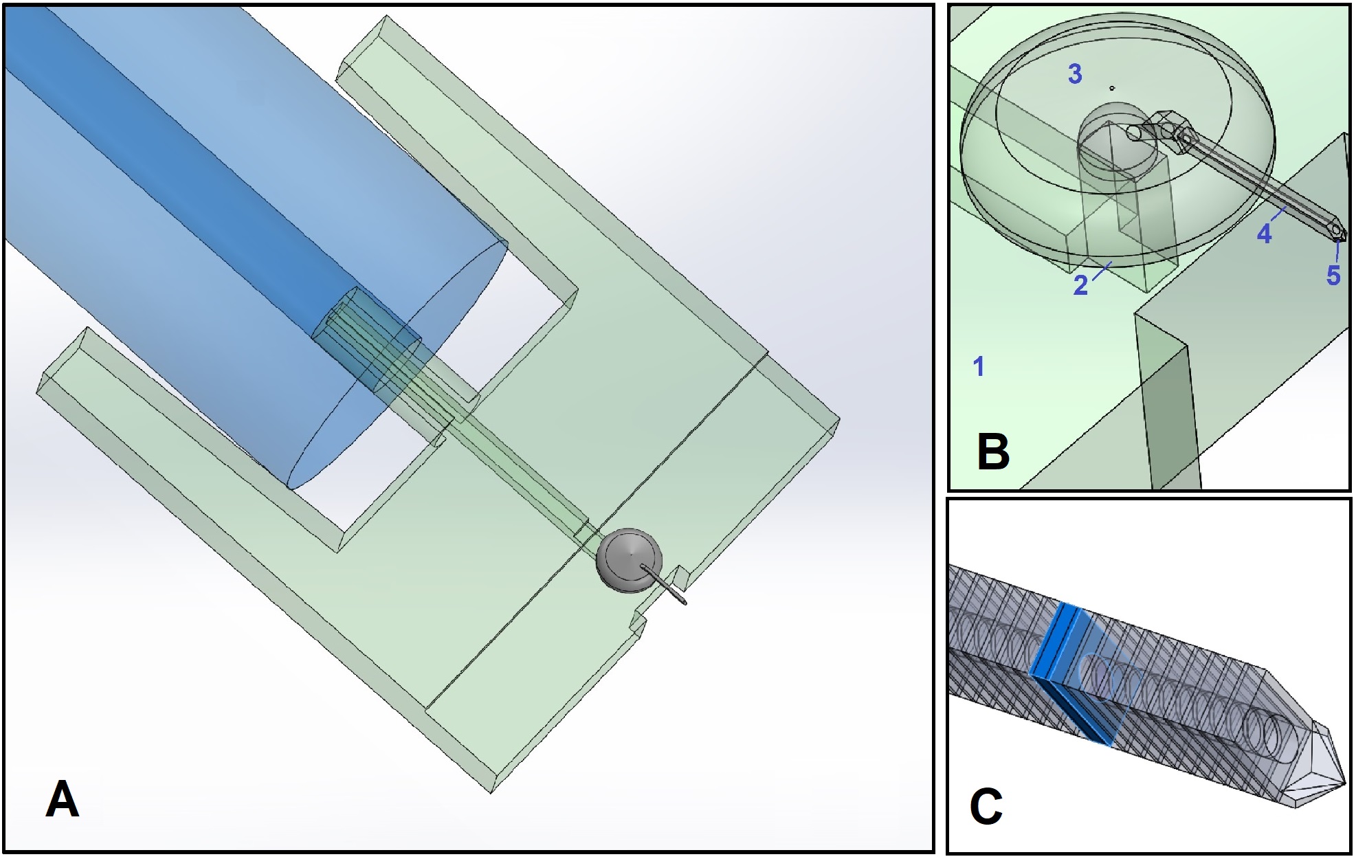
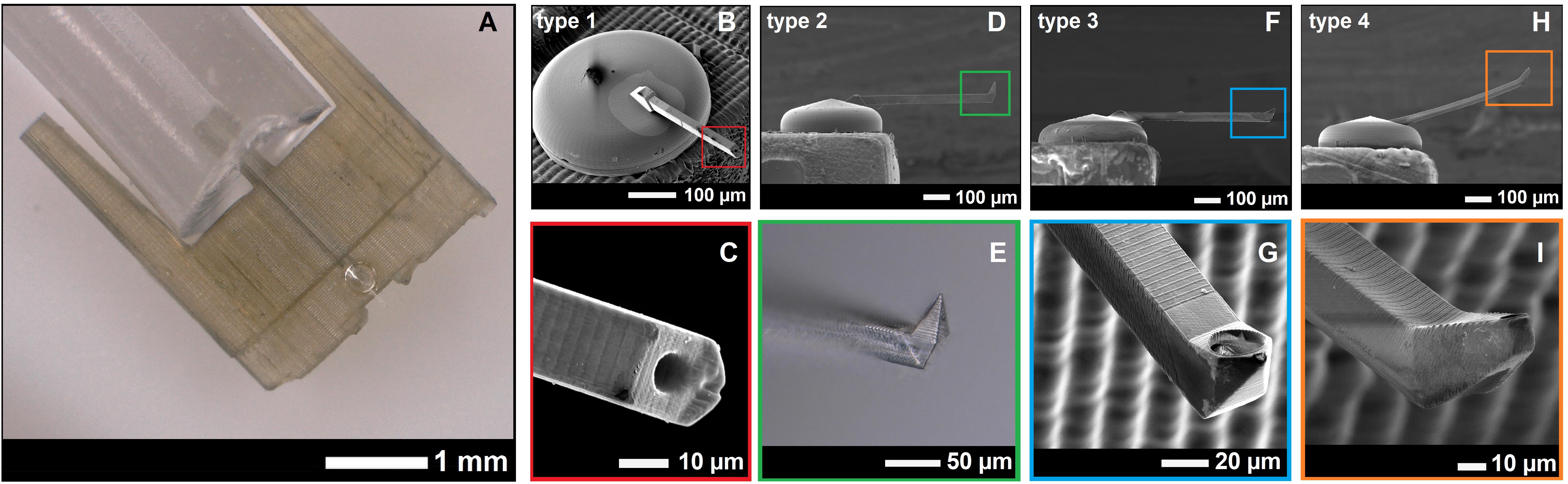
To analyse diseases, researchers often perform single-cell biopsies using a Microfluidic Atomic Force Microscope (AFM) and a tailor-made cantilever. An AFM is a special tool with a sharp needle mounted on a flexible cantilever. During a single-cell biopsy, the needle meticulously penetrates the cell in order to reach the desired location. Developing prototypes for this is frequently time-consuming and costly as a result of the complicated production process. Ghatkesar’s 3D technique enables researchers to develop prototypes for single-cell biopsies much more easily and quickly. This can have huge benefits for medical research. Indeed, Murali Ghatkesar would like to use this new technique to conduct further research on muscle dystrophy in people with paralysis on one side of the face, for example.
Murali Ghatkesar conducted his research in cooperation with Leiden University and his own research team.
Read the publication Multiscale 3D-printing of Microfluidic AFM Cantilevers
Read more about Murali Ghatkesar’s research in
“Quantifying the nanoworld: new tools for nanoscientists”