Advanced microscopy to understand life and fight disease
On 20 February, NL-BioImaging (NL-BI) received national funding of 25 million euro, of which 15 million by NWO. The funding supports the consortium in becoming the national advanced light microscopy infrastructure providing coordinated access to the Netherlands’ best imaging technology and analysis platforms. NL-BI is a multi-sited collaboration of all 18 Dutch universities, medical academic centres and research institutes.
In the NL-BI consortium, scientists from all Dutch academic research centres will together develop and integrate state-of-the-art microscopy with technologies and services in different nodes. This will enable access for all scientists to revolutionise fundamental insights into the building blocks of life, enable scientific breakthroughs, and advance applications towards society for overcoming life-threatening disease, including cancer, metabolic, cardiovascular, and neurodegenerative disorders.
Physicists Jacob Hoogenboom and Bernd Rieger from the Imaging Physics department at TU Delft will contribute to the NL-BI nodes on correlative light and electron microscopy (CLEM) and smart microscopy.
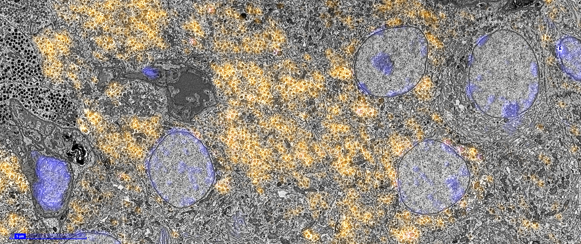
''In the CLEM node, we can study the whereabouts of proteins in cells, tissue, organs, and organisms'', Hoogenboom explains. ''We develop instruments to allow large scale correlation between fluorescence and electron microscopy in high resolution and in 3D. With fluorescence microscopy, we can visualise biological molecules, proteins, and their spatial distribution. Electron microscopy allows us to look at the biological structure.''
In smart microscopy, Rieger and co-researchers will develop software and instrumentation with which a sample can be analysed unsupervised: the microscope is self-steering. Rieger: “Software recognises interesting, possibly rare events on the fly and adapts the imaging protocol to optimally retrieve data from these events. A smart microscope enables much higher throughput of biological samples and allows to find rare events in higher numbers more quickly, such that biologists can draw conclusions on a wider statistical basis.”
Life and disease
Since Antoni van Leeuwenhoek first steps in life sciences using microscopy, innovative microscopy has become essential for life sciences with exciting recent progresses in biosensors, microscopic resolution, and three-dimensional imaging (3D) imaging. Coupled with contemporary advancements in quantitative image analysis and the capacity to leverage Artificial Intelligence (AI) for mining large amounts of image data, scientists have reached a significant milestone in their ability to detect and manipulate the behaviour of molecules and live cells in organoids, tissues, and small animals, allowing us to gain a deeper understanding of the fundamentals of life and disease.
Overcoming challenges
However, the required investments in high-tech instrumentation, as well as challenges in the development of fluorescent probes and data-analysis tools, have become impossible to sustain for individual life science laboratories or even individual microscopy facilities. NL-BioImaging (NL-BI) aims to overcome these challenges by jointly bridging technology gaps and offering access to advance functional imaging in complex systems at all scales.
Read the full press release at Microscopie.nl

