
We have 2 new student projects available at the Carroll Lab where we use light and electron microscopy to study how the brain develops. Our goal is to develop tools to image synapse function and structure in living zebrafish embryos, and connect this to nanoscale maps made using correlative photon and electron techniques.
- 3D Reconstructions of individual neurons
Neurons in the brain are the most complex cell types in the entire body. We use sparse fluorescence labelling techniques to visualize individual neurons developing in the zebrafish. Neurons can then be imaged by light microscopy techniques, like confocal laser scanning and light sheet microscopy. From images of the same neuron on different days of development, we want to quantify changes in the shape of the neuron. For this project, we are using the latest generation of 3D neuron tracking and volume rendering softwares.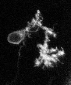
- Tissue staining for light and electron microscopy
While electron microscopy can obtain spatial resolution of a few nanometers, light microscopy offers more options to obtain information about biological content of tissue. The two techniques can be combined in correlative light-electron microscopy, but some trade-offs exist: standard stains to obtain the best contrast in electron microscopy typically destroy fluorescence for light microscopy. We are testing staining protocols in zebrafish tissue to optimize visualization of synaptic structures with both light and electron imaging modalities.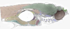
More information or addtional questions? Please contact Dr. Elizabeth Carroll.