Clever Delft trick enables 20 times faster imaging with electron microscopy
Researchers at TU Delft have expanded upon a clever trick, thereby increasing the speed of electron microscope imaging by a factor of twenty. A simple adjustment is all that is needed: applying a voltage to the specimen holder. Through this simple intervention, a specimen that would normally take an electron microscope a week to image can now be inspected in a single night or one working day.
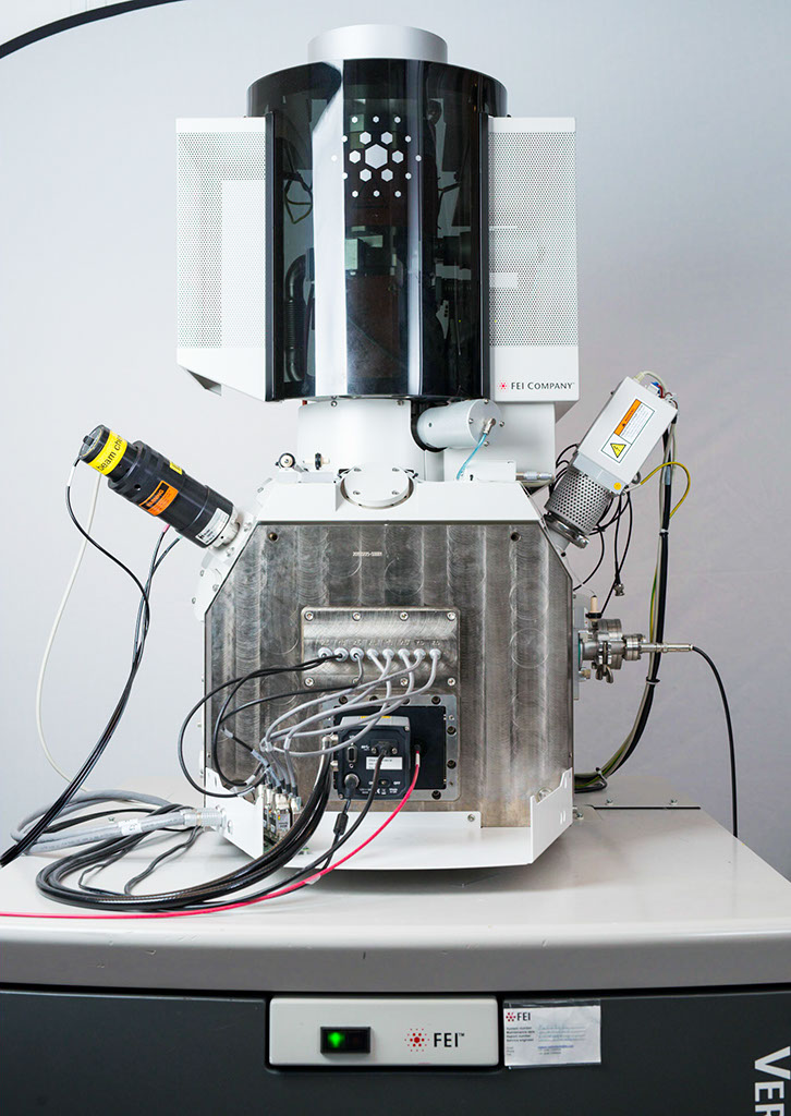
Electron microscopes are unparalleled when it comes to imaging at the very smallest scale. Unlike an optical microscope that captures light particles, the scanning electron microscope (SEM) shoots an electron beam at the specimen—for example, a thin slice of tissue. The electrons in the beam scatter in the tissue, whereupon the scattered electrons are captured by a sensor. Then a computer creates an image based on how many electrons are scattered at each position the beam scans.
Painstaking task
Electron microscopes are capable of magnifying objects up to a million times, enabling you to examine the structure of the tissue or other material at almost molecular level. But because the device works in such detail, it is a truly painstaking task to map out small objects. “As part of research into diabetes, for example, we are working together with researchers from UMC Groningen to create images of the insulin-producing islets of Langerhans in the pancreas," says TU Delft researcher Jacob Hoogenboom. “To create a full image of just one slice of tissue of a pancreas from a rat takes around a week's worth of measurements.”
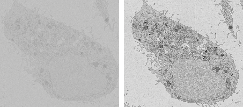
Hoogenboom's research group, which is working on improving optical and electron microscopes, has now come up with a smart trick to speed up this process by a factor of twenty. “Applying a voltage to the specimen holder enables us to slow down the incoming electrons while simultaneously accelerating the outgoing electrons," he explains. “This acceleration means the electrons hit the detector with more energy so they generate more signal thereby overwhelming electronics noise and shot noise and enabling the device to measure more efficiently.”

Dr. ir. Jacob Hoogenboom
Potential
The researchers came up with this idea through a different, fundamental, research project. In this project, they are trying to work out what chemical reactions the electrons enter into with the materials they are scanning, at different energies. “The aim is to limit these reactions as much as possible, because you don't want what you are scanning to change while you are scanning it," says Hoogenboom. “To adjust the energy of the electrons, we also experimented with applying an electric potential to the specimen. We were discussing this among ourselves when we realised that we could also use such a potential to save time while creating an image.”
The great thing about this TU Delft trick is that it doesn't require any complicated adjustments: anyone with a scanning electron microscope can use it. Hoogenboom: “In most electron microscopes it is already possible to apply a voltage to the specimen holder. Normally people use this option to separate high-energy scattered electrons from those with low energy, so you are only left with the useful signal. But until now no one had realised how much more quickly you are able to image your specimen.”
More information
‘Optimization of negative stage bias potential for faster imaging in large-scale electron microscopy’, R. Lane, Y. Vos, A.H.G. Wolters, L. van Kessel, S. Elisa Chen, N. Liv, J. Klumperman, B.N.G. Giepmans, J.P. Hoogenboom, Journal of Structural Biology