3-in-1 microscope shows researchers the way to proteins
Physicists from TU Delft have developed a 3-in-1 microscope where a light beam, electron beam and ion beam work together to precisely cut out specific slices from biological samples. These slices are indispensable for biomolecular research into new generations of medicines. The invention was published in the journal eLife today.
Biomolecules in the cell
The foundations of health and disease can be traced back to the functioning or non-functioning of biomolecules in the cell, such as proteins. Research into these molecules is essential for the development of medicines. In 2017, the Nobel Prize in Chemistry was awarded to the inventors of a microscopy technique that can reveal the smallest and most complex biomolecules.
This so-called cryo-transmission electron microscopy (cryo-TEM) uses an electron beam to image individual biomolecules in the cell, down to almost the atomic scale. To do this, the cell must be rapidly frozen to cryogenic temperatures (typically –170°C) and a wafer-thin slice containing the targeted biomolecule has to be cut out of it. In practice it is very difficult to cut exactly in the right place.
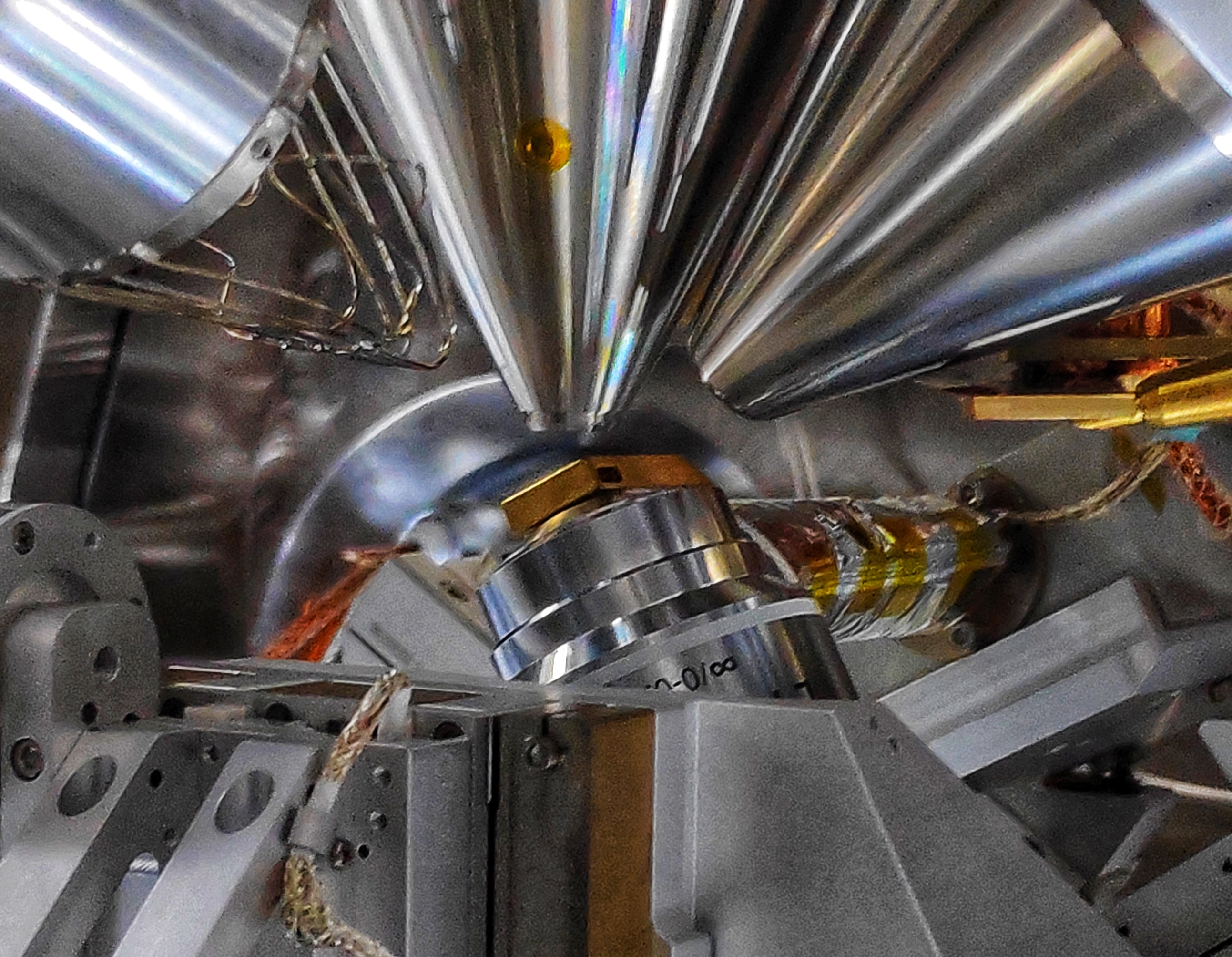
Three bundels
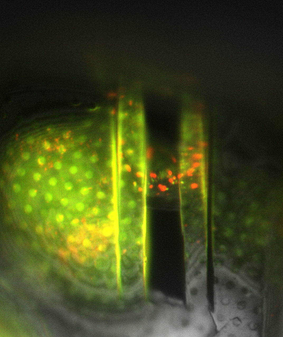
PhD candidate Daan Boltje has now developed a technique for simultaneously examining the cell using three microscopy techniques. By attaching a fluorescent label to the target biomolecule, it literally lights up, after which a slice is cut out around the location of the fluorescence, after which it can be further investigated with cryo-TEM. The use of cryo-TEM for research into proteins and other biomolecules has exploded in recent years. Boltjes' invention makes such cryo-TEM research easier and more precise.
To make this possible, Boltje simultaneously focused three beams on a biological sample: a light beam to light up the fluorescent dyes, an electron beam to zoom in on the nanometer scale, and an ion beam to cut out a thin slice.
Technical achievement
Lead researcher Jacob Hoogenboom: “We cut a 0.1 micrometer thick slice from a cell of 10,000 cubic micrometers in exactly the right place. In vacuum and at –170°C. Technically, that's quite an achievement." Previously it was necessary to examine the sample in separate microscopes, after which researchers had to superimpose the data from these microscopes. Alternatively, they cut out a ‘random’ set of slices in the hope that one of them contained the wanted biomolecule.
Boltje, who is an industrial PhD candidate at both TU Delft and microscopy company Delmic, developed the technique in collaboration with cell biologists from the Netherlands, Germany, Australia and the US, who as end users will immediately apply the system for their research into proteins. “I am especially proud of how easy to use the system is. You see the fluorescence on your screen, you navigate towards it, and with the push of a button the microscope cuts out a slice there.”
Delmic has been working on combining light and electron microscopy for some time now. Hoogenboom: “Daan has shown how light, ions, electrons and cryogenic cooling can come together in one vacuum system. Three prototypes will now be used by other groups. Our challenge is to further develop the technology and perhaps bring it to market in the long term.”

