From light spots to supersharp images
Making detailed 3D images of proteins in living cells with a special light microscope, without damaging those cells. That is what Sjoerd Stallinga, winner of an ERC Advanced grant worth 2.3 million euros, wants to achieve. In order to do so he is going to scan samples nanometer by nanometer using a sophisticated 3D light pattern in an approach that requires extensive collaboration between different disciplines.
Nowadays nobody uses their own eyes to look at a sample under a light microscope
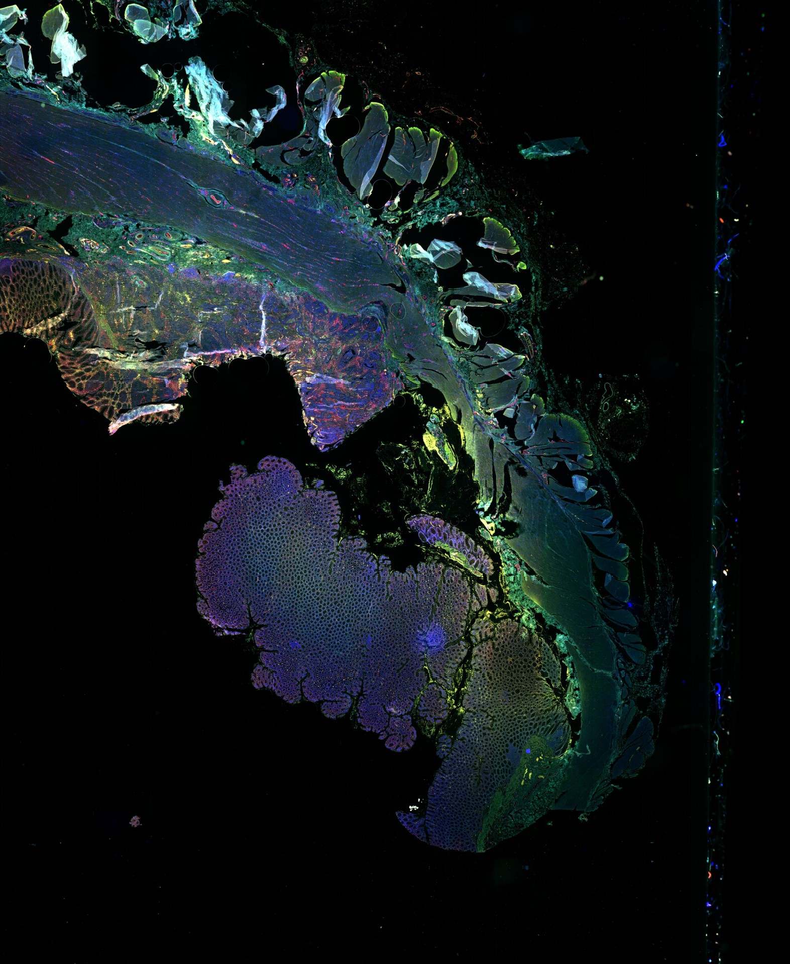
Computational imaging is what the world of microscopy is all about these days. Without a computer there is no image. “Nowadays nobody uses their own eyes to look at a sample under a light microscope,” says Professor Sjoerd Stallinga, chairman of the Imaging Physics Department. Doing so would mean you only see large structures, such as the protein factories of a cell, the so-called ribosomes. If you also want to pinpoint the location of individual proteins in a cell, or map how protein complexes are assembled, a smart interaction between the microscopic techniques used and the image processing software is essential.
Reinforcing each other
Stallinga’s prize-winning research plan calls for collaboration between four very different disciplines, namely engineering optics, computer science, biochemistry and biology. “Choosing a multidisciplinary approach like this works because it leads to all kinds of innovative things.”
“Research groups within my department also reinforce each other. Technology developed for one imaging technique is often equally valuable for improving completely different imaging techniques,” Stallinga continues. He regards receiving this ERC grant, which is the largest European prize for an individual researcher, not only as recognition of his own work, but also of all the people around him. “If you walk up the corridor here, you’ll see that all my colleagues, just like myself, team up with researchers who use their techniques, as well as with companies that sell the technologies they work on.”
Fluorescent labels
His own specialty is super-resolution microscopy. This Nobel Prize winning technique already enables zooming in to the level where you can see individual protein complexes. However, if you also want to look at individual proteins and at the structure of protein complexes in more detail, you need to go a step further. “In order to do that you need a resolution of 1 to 5 nanometers, which is about five times more detailed than images produced by standard super-resolution microscopy,” Stallinga explains.
That resolution has already been feasible with electron microscopy for a long time. However, that kind of microscopy not only requires preparatory measures that eventually kill a cell, but also makes it impossible to use a range of fluorescent labels to make specific proteins identifiable, let alone create 3D images and that is something Stallinga has set his sights on.
Twinkling stars
Nobel Prize winners Eric Betzig, Stefan Hell and William Moerner discovered that you can get more accurate images with light microscopy if you attach fluorescent labels to proteins that emit light only part of the time, like blinking lights. Two proteins that are close to each other can then still be distinguished from each other if they do not emit light at the same time. If both were to emit light at the same time, only one combined light spot would be visible, rather than two separate ones. If only one light spot is visible at a time, the location of each protein can be estimated from the centre of that light spot.
Compare it to a night sky full of twinkling stars, only in our case the stars don’t give off any light for 99 percent of the time.
“Compare it to a night sky full of twinkling stars,” the Delft-based physicist suggests. “Only in our case the stars don’t give off any light for 99 percent of the time.” This means that if you have a sample with a fluorescent label attached to ten thousand proteins, at any specific moment only about a hundred of those labels will actually emit light and those hundred will be randomly distributed across the sample. Each picture will then depict different proteins. Superimposing all the detected protein positions will produce a complete and supersharp image.
Lines and dots
In order to make images even more accurate and make 3D images at the same time, Stallinga plans to illuminate a sample with a 3D light pattern with dark and light parts. “We want to make a pattern of stripes and dots of light in three dimensions.” He then wants to use that to make a kind of scan in which the brightness of a spot of light increases and decreases as a line or dot of light slides by. In this way Stallinga expects to achieve a higher resolution than with uniform illumination. Another advantage is that this can be done with comparatively little light, meaning a minimum of damage to the sample, which is largely transparent. “Uniform exposure requires so much light that your own skin would turn red.”
Pathologists
For the next couple of years Stallinga’s goal is to double the resolution in 3D samples in the x and y directions. In the z direction he is even going to strive to gain a sixfold increase. “Often the resolution in the z direction is currently three times worse than in the x and y directions,” he explains. He also wants to achieve this gain across larger volumes, say 100 by 100 by 100 micrometers. That is comparable with the thickness of a hair. ”But for us, that’s pretty big.”
If his research progresses well, it will give the medical sector all kinds of new opportunities. Stallinga is aware that imaging techniques already play an incredibly important role in healthcare and improving super-resolution microscopy can enhance that role even more, for example when used in the search for drug targets. Another example is neuroimaging which uses light microscopy to examine the interaction of neurons and to investigate diseases such as Alzheimer’s.
He also expects spin-offs in other areas. “We’re also going to develop new techniques for image processing and analysis. If we invent generic solutions in this field, they may also be useful to pathologists, for example by making it easier to detect abnormalities in tissues.”
