Organ-on-a-Chip: Interfacing with living tissue
“A what on a chip?” is what most people ask with disbelief when they hear for the first time of the new and very exciting research area “Organ on a Chip”, according to Prof Dr Ronald Dekker. The topic has nothing to do with Science Fiction, but everything with a very surprising encounter between two completely different research fields: Micro-fabrication and Stem-Cell Biology! The final goal is to build simplified, but very well controlled models of human organs and diseases
One of the principles of science is to translate a complex problem into a simple model. Once the simple model is fully understood, more complexity can be added. If you want to study the formation of rain for example, you can take a beaker and carefully control the: temperature, humidity, pressure and particle density. It is here that medical sciences so far have had a problem: the human body is an extremely complex system, with many factors interacting of some which are even not known. It is simply not possible to take for instance a lung out of a person to study what is going on in detail. Organ-on-a-Chip is a new research field which has the ambition to generate breakthrough solutions for exactly this problem.
Heart-on-a-chip
In the Else Kooi Laboratory we have been working already for a number of years on an Organ-on-a-Chip model for the human heart, which can actually mimic the human heart during exercise. The primary goal of the model is to screen new drugs at a very early stage of their development for possible side-effects which can cause lethal arrhythmias. In the model a piece of human heart muscle tissue is grown on a thin stretchable membrane containing electrodes. The electrodes register the electrical activity of the cells while they are subjected to the drug compound. By stretching the membrane it is possible to simulate physical exercise which is an important factor in the occurrence of these arrhythmias. The project is done in close cooperation with the group of prof. Christine Mummery at the LUMC in Leiden which has a world renowned expertise in the differentiation of human stem cells into heart muscle cells. The technology platform we developed – coined Cytostretch – has the unique property that it allows for electrical characterization of cells while they are mechanically stretched. It is the intention to use the Cytostretch platform also as a model for cardiac diseases like: heart failure, hypertrophic cardiomyopathy, and congenital cardiac arrhythmias. Also other diseases involving electrically active cells such as: muscular dystrophies, brain diseases, peripheral nerve cell diseases, and pain syndromes, can be studied with the Cytostretch platform.
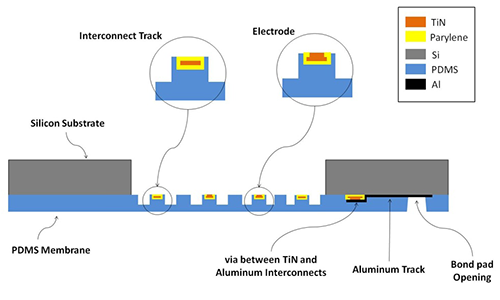
The Cytostretch device is used to measure the electrical activity of cells under stretch. It consist of an array of stretchable electrodes embedded in a silicone membrane.
Live view