Cytoskeletal crosstalk during cell division
Ilina Bareja
Collaborators: Gijsje Koenderink (TU Delft), Zdenek Lansky and Marcus Braun (BIOCEV Research centre, Prague)
Microtubules and actin are known to play crucial roles for the proper functioning of cells, and the two types of cytoskeletal filaments have been studied extensively separately, both in- vitro and in-vivo. However, inside the cell these two filament types work together in a highly regulated fashion, and only in the past few years has the focus shifted to study these components together. Such crosstalk processes involve both indirect (cell signaling) and direct (crosslinking proteins) communication between the cytoskeleton. One striking example of this crosstalk occurs during the process of animal cell division- whereby microtubules emanating from centrosomes can help in positioning the cytokinetic ring (made of actin filaments) in the middle. The actin cortex on the other hand helps in correctly positioning the centrosomes.
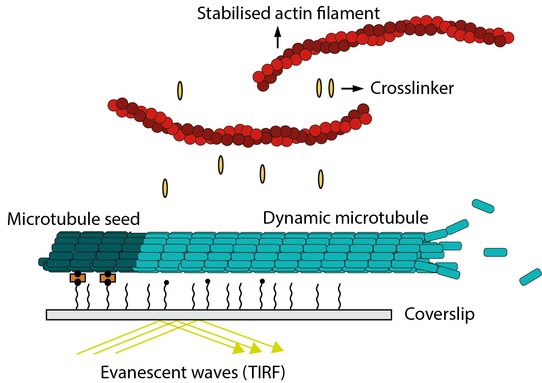
Here, we focus on studying the direct crosstalk between actin and microtubules using proteins which potentially can act as direct cytoskeletal crosslinkers during animal cell division. For this, we use a bottom up approach to visualise fluorescently labelled microtubules, actin filaments and crosslinkers inside channels.