In vitro reconstitution of the microtubule-kinetochore coupling
Vladimir Volkov and Ali Nick Maleki
Collaborators: Prof. Andrea Musacchio, Max Planck Institute of Molecular Physiology in Dortmund, Germany.
Cell division is one of the most fundamental processes that aims to distribute two copies of genome to two daughter cells. The physical movement and separation of chromosomes relies on the coupling of dynamic microtubule ends to the kinetochores, multi-protein complexes forming on the centromeric regions of chromosomes.
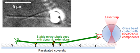
We use single-molecule fluorescence and optical trapping to reconstitute the dynamic coupling of microtubule tips with a simplistic experimental model of a human kinetochore. Using this bottom-up approach we probe forces exerted by individual tubulin protofilaments and elucidate the mechanisms allowing the kinetochores to tune microtubule dynamics.